Images and Scans
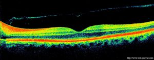
Optical Coherence Tomography (OCT)
OCT is an imaging technique to capture micrometer resolution, and two and three dimensional images of the retina and anterior segment. OCT provides a straightforward method of assessing cellular organisation, axonal thickness in glaucoma, macular degeneration, diabetic macular edema, multiple sclerosis and other eye diseases or systemic pathologies which have ocular signs.
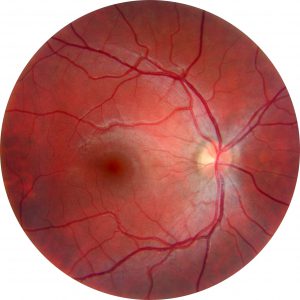
Digital Retinal Photography
A scan of the retina to confirm a healthy eye or detect the presence of disease. The image gives the Optometrist a more detailed view of the retina than they can achieve by other means. By comparing images from previous visits, the Optometrist will easily be able to detect slight changes over time and be able to detect and help prevent potential macular degeneration, glaucoma, retinal tears, optic strokes and much more.
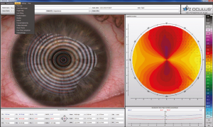
Oculus Keratograph
Keratograph 5M technology is a revolution in corneal topography and dry eye analysis. The high-resolution colour camera and the integrated magnification changer offer a new perspective on the tear film assessment procedure.
The Keratograph 5M measures corneal topography precisely. The built-in real keratometer and automatic measurement activation guarantee perfect reproducibility of K values. Data is acquired by noncontact measurement, automatically analysed and shown in comprehensive presentation formats.
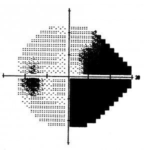
Visual Field Analysis
The visual field test is a subjective measure of central and peripheral vision and is used to diagnose, determine the severity of, and monitor glaucoma. It evaluates vision loss due to glaucoma, damage to the visual pathways of the brain, and other optic nerve diseases. When glaucoma is diagnosed the visual field data is used to determine the severity of disease. This staging information is useful in choosing a target intraocular pressure and determining follow-up.